Diffuse Optics Technologies
Diffuse optics devices offer portable fiber-coupled instrumentation for non-invasive estimation of deep tissue hemodynamics (1-2 cm below surface) at few spatial positions. Illumination and detection near-infrared light (~650-850 nm) is realized via custom fiber optic probes with source and detector positions 2-3 cm away on the tissue surface. Hybrid diffuse optical devices utilize two complementary technologies for quantitative measurement of tissue blood oxygenation and tissue blood flow.
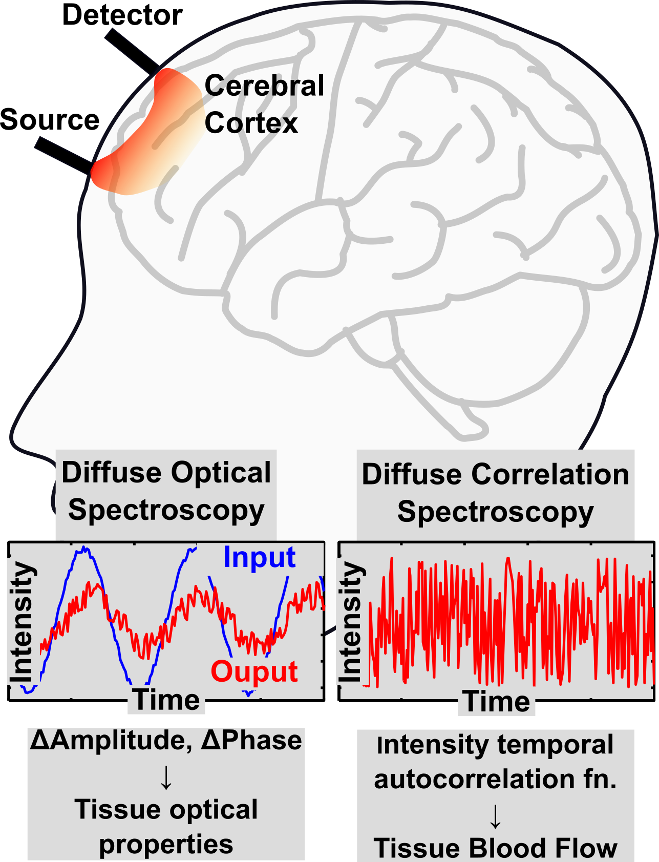
Diffuse Optical/Near Infrared Spectroscopy (DOS/NIRS):
Diffuse Optical Spectroscopy is used to estimate concentrations of oxygenated/deoxygenated hemoglobin (and hence tissue oxygen saturation). Several components of tissue (e.g., oxy- or deoxy- hemoglobin, lipids, water) absorb energy from light at different wavelengths, in quantities that are proportional to their concentrations. DOS aims to estimate these concentrations by measuring the differential absorption of multispectral light by tissue.
The most common and simplest implementation of DOS is Continuous Wave Near-Infrared Spectroscopy (CW-NIRS). Here, multi-wavelength near-infrared light of constant (i.e., continuous) intensity is used to illuminate the tissue, and the intensity of diffuse reflected light is recorded. The absorbance/attenuation is then calculated at multiple wavelengths, and the modified Beer-Lambert law is used to estimate changes in oxygenated and deoxygenated hemoglobin. CW-NIRS offers a fast and convenient method, but is limited to measurements of changes in hemoglobin concentrations.
More quantitative approaches utilize intensity modulated light to derive both tissue absorption and tissue scattering properties. Simultaneous measurement of these optical properties enables separation of attenuation of light due to absorption or scattering, and estimation of baseline tissue hemoglobin concentrations.
-
Frequency-Domain Diffuse Optical Spectroscopy (FD-DOS):
In Frequency Domain Diffuse Optical Spectroscopy (FD-DOS), the intensity of illumination light is modulated sinusoidally (~100 MHz), and the phase and amplitude changes of light propagated through tissue is measured at different source-detector separations. When combined with a photon diffusion model, the simultaneously measured amplitude and phase changes permit quantification of both tissue absorption and scattering coefficients. -
Time-Domain Diffuse Optical Spectroscopy (TD-DOS):
Time Domain Diffuse Optical Spectroscopy (FD-DOS) illuminates the tissue with light pulses of very short durations (~100 ps), and measures the time delayed and broadened light that emerges at detector positons. The shape of the detected pulse is used to estimate tissue scattering and absorption properties.
Diffuse Correlation Spectroscopy (DCS):
In contrast to DOS, Diffuse Correlation Spectroscopy (DCS) is a realtively new and qualitiatively different technique used to measure tissue blood flow. DCS illuminates the tissue with highly coherent light from a single-mode laser; light scatters via different path lengths through the tissue leading to interference (i.e., speckle) at the detector positions. Moving partices (e.g., red blood cells) in the sampling volume modulate/change these paths causing the detected intensity to flucuate in time. DCS measures the scale of these fluctuations by converting the intensity fluctuations to intensity temporal autocorrelation functions, and estimates blood flow by fitting the autocorrelation functions to a correlation diffusion model.
(See Diffuse correlation spectroscopies for non-invasive, micro-vascular cerebral blood flow measurement, by Durduran & Yodh, Neuroimage (2014) for a comprehensive review of DCS techniques)
Back to Research Topics
Relevant Publications
Wang D, Parthasarathy AB*, Baker WB, Gannon K, Kavuri V, Ko TS, Schenkel S, Li Z, Li Z, Mullen TM, Detre JA, and Yodh AG (2016), “Fast blood flow monitoring in deep tissues with realtime software correlators”, Biomedical Optics Express, 7 (3), 776-797. PMID: 27231588 PMCID: PMC4866455. (PDF)
Li Z, Baker WB, Parthasarathy AB, Ko TS, Wang D, Schenkel S, Durduran T, Li G, and Yodh AG (2015). “Calibration of diffuse correlation spectroscopy blood flow index with venous-occlusion diffuse optical spectroscopy in skeletal muscle”, Journal of Biomedical Optics, 20 (12), 125005. PMID: 26720870 PMCID: PMC4688416. (PDF)
Baker WB, Parthasarathy AB, Ko T, Busch DR, Abramson K, Mesquita RC, Durduran T, Greenberg JH, Kung D and Yodh AG (2015). “Probe pressure modulation algorithm reduces extra-cerebral contamination in optical measurements of cerebral hemodynamics”, Neurophotonics, 2 (3), 035004 PMID: 26301255 PMCID: PMC4524732.(PDF)
Baker WB, Parthasarathy AB, Busch DR, Mesquita RC, Greenberg JH, and Yodh AG (2014). “Modified Beer-Lambert law for blood flow”, Biomedical Optics Express, 5 (11), 4053-75 PMID: 25426330 PMCID: PMC4242038.(PDF)
Buckley EM, Parthasarathy AB, P Ellen Grant, Yodh AG, and Franceschini MA (2014). “Diffuse correlation spectroscopy for measurement of cerebral blood flow: future prospects”, Neurophotonics, 1 (1), 011009, PMID: 25593978 PMCID: PMC4292799.(PDF)